The PuLSSE Lab is located on the first floor of the Nursing & Science Building at Rutgers University-Camden. For directions, please click here.
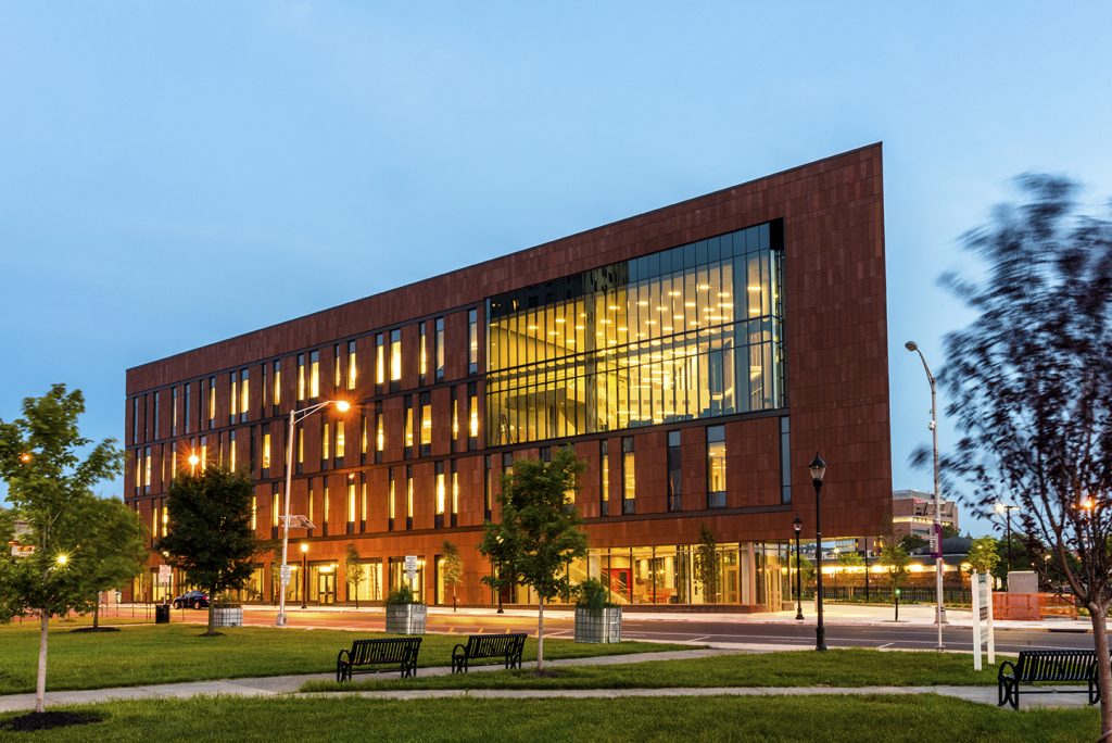
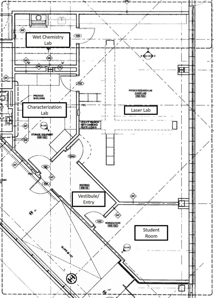
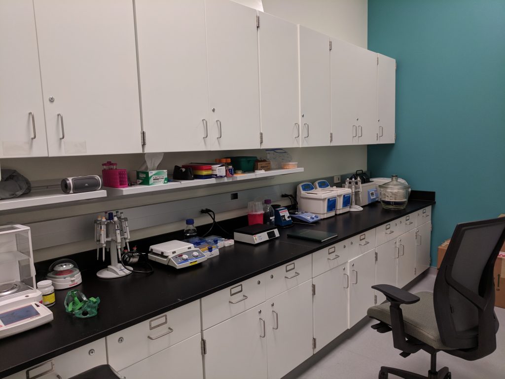
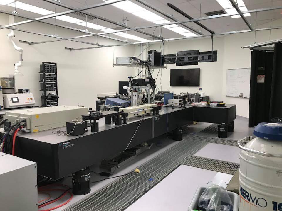
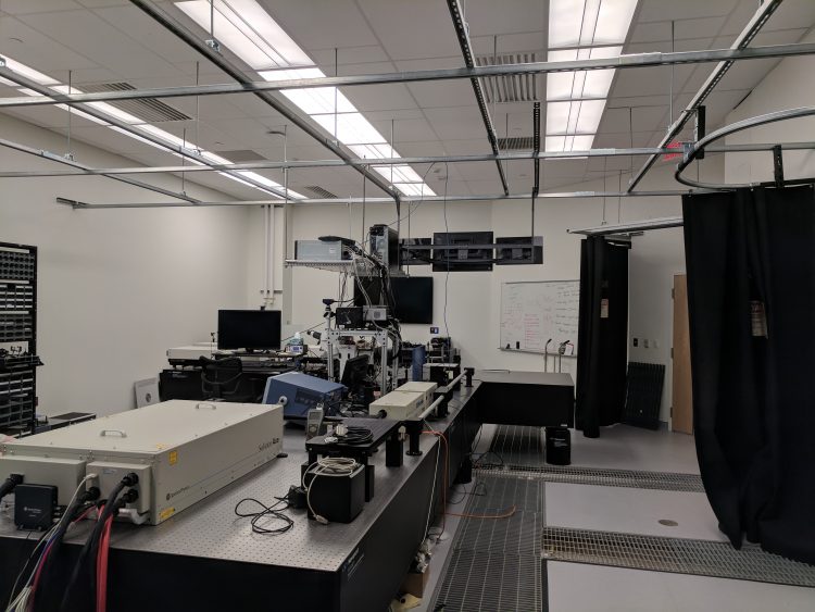
Our team shares a common experimental laboratory that was opened in the Fall of 2017. Approximately 2000 square feet of space was custom built to suit the unique needs of the PuLSSE Group. Given the urban setting that this building is in, several steps were taken to remediate vibrations. The lab is located on the ground floor and was built on an isolated foundation. All vibration sensitive equipment is placed on a custom designed 144 square foot Newport optical table. The space is divided into five separate rooms: a wet chemistry lab, characterization lab, laser lab, student workspace, and an isolated entryway for addition laser safety. The laboratory is secured with card access, and building access is controlled by on-site security.
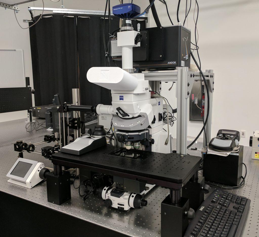
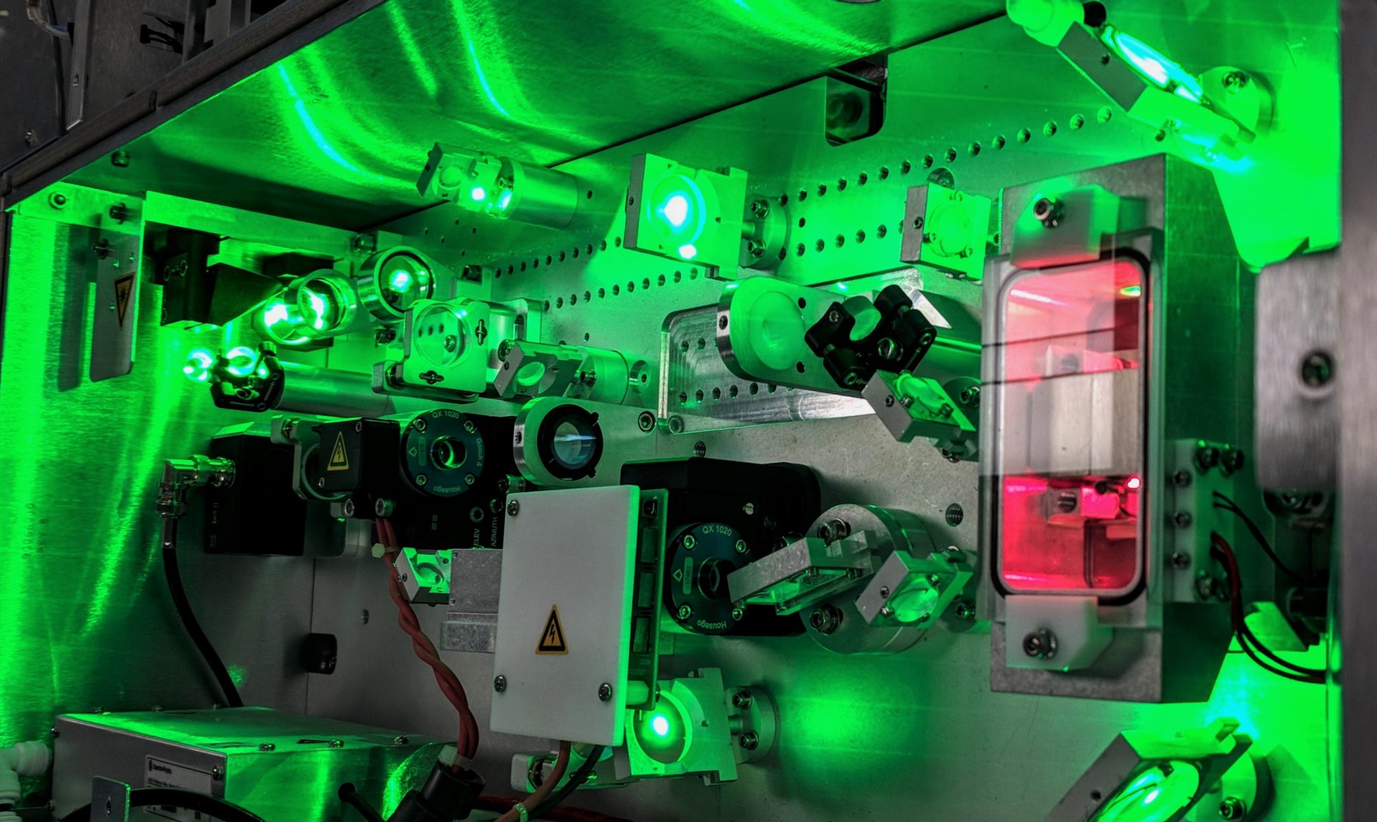
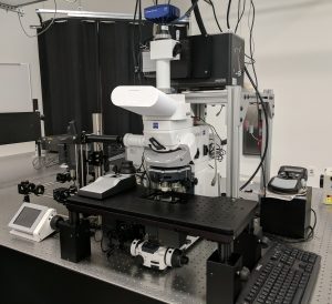
We recently acquired a unique ultrafast laser system integrated with a research-grade optical microscope funded through NSF-MRI award #1531783. The laser is a femtosecond Ti:sapphire system pumping an automated optical parameter amplifier with sum-frequency and harmonic generation stages that allow for a broad selection of wavelengths (240-2400 nm). The selected output is steered into a fixed-stage upright microscope whereby the optical and laser paths are made collinear via dichroic mirrors. Additionally, a 785 nm CW diode laser has also been combined into the optical path to facilitate optical tweezing.
The instrumentation is meant to dramatically expand the scope and significance of research being conducted in southern New Jersey in the areas of: ultrafast laser ablation/machining, membrane photoporation/phototransfection, plasmonic nanoparticle scattering, and multiphoton absorption polymerization.
Key components:
• Spectra Physics Solstice ACE 100F1K Ti:Sapphire laser (800 nm fundamental, 120 fs, 6 mJ, 1000 Hz) + 3rd Harmonic generation and TPR-TOPAS for generation of light from 240 nm-2400 nm
•Zeiss AxioExaminer Z1 fixed stage upright microscope – can perform fluorescent, darkfield, and phase contrast imaging.
•Andor Kymera193i Spectrograph equipped with dual exit ports to which we have attached an iStar iCCD and a Neo5.5 sCMOS cameras.
Beamtime with the system is available to both internal and external users with special priority given to investigators from institutions located in South Jersey. Interested parties should contact Dr. Sean O’Malley to discuss whether the capabilities of the system can meet the needs of your research project.
Additional equipment and instrumentation:
- Exspla NL-303 ns Nd:YAG + OPO. Tunable from 400 nm to 2600 nm and with a 4 ns pulse duration (550 mJ at 1064 nm, 200 mJ 532 nm, and 70 mJ 266 nm)
- Exspla Nl 201 Nd:YAG (532 nm, 0.3 mJ, 7ns) laser
- Exspla PL-2241 2. 25 ps Nd:YAG (250 Hz, 5 mJ 1064, 2 mJ 532, 0.7 mJ 266)
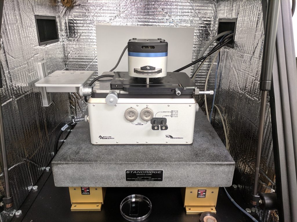
- AFM – an MFP-3D Asylum atomic force microscope
- Olympus imaging microscope for use with AFM
- Raman spectrometer with Princeton Instruments Spectrpro-2500 and PIXIS:100B eXcelon Digital CCD Camera System. Can measure fluorescence and Raman spectra from 100-4000 cm-1, Wavelength of operation – 532 and 785 nm
- Electronics – 1GHz & 300 MHz HP oscilloscope, HP 3562a spectrum analyzer (64 µHz – 150 kHz), Stanford 830 lock-in, detectors (MCT, etc.)
- Vacuum chamber for deposition, pumps, valves, thermocouple, convectron, etc.
- Nikon stereo optical microscope – designed for wire bonding, soldering, etc
- Denton dual source evaporator
- Maktek TM 400 and QPOD- quartz crystal microbalance
- Malvern Zetasizer ZS 90 DLS unit (13o, 173o, and zeta potential)
- Cary Eclipse Fluorimeter
- Cary 60 Uv-VIS spectrometer
- Bruker ALPHA FTIR Spectrometer
- Flashforge 3D printer, dual extruder, Dremel 3D Printers
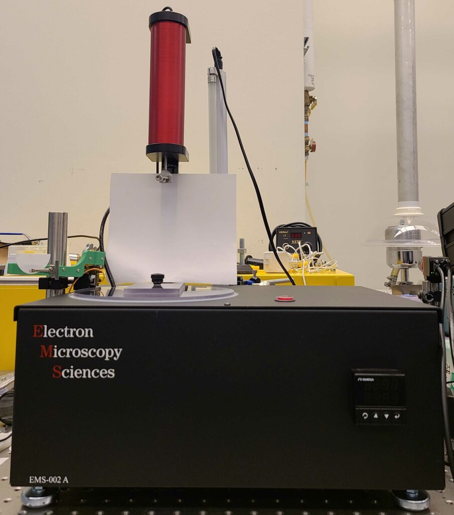
- Electron Microscopy Sciences Plunge Freezer (EMS-002) for vitrification
- Mettler Toledo MX5 Microbalance
- Denton Desk II Sputter Coater
Please contact Dr. Sean O’Malley or Dr. Julianne Griepenburg if our equipment can assist you with your research needs.